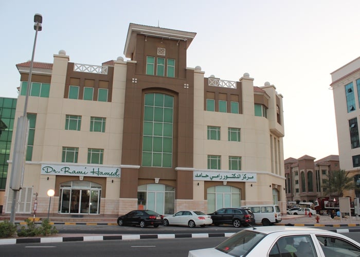
Joint Pain in Childhood at Pediatric Clinic DRHC Dubai
- The history and physical examination are key to establishing the correct diagnosis of joint pain in childhood, and according to that, the Doctor will ask for laboratory and radiologic examination for final diagnosis and appropriate treatment.
- It’s important to know the nature of pain, duration, frequency, stiffness, and severity. The timing, how many joints are involved and family history of rheumatoid disease, and if there is any other symptoms such as fever, weight loss, fatigue, gastrointestinal symptoms (diarrhea, blood in stool), signs of previous urinary tract infection, tonsillitis, skin rash and any symptoms of inflammation (redness, effusion, warmth) or just joint pain without signs of inflammation, and any precipitating factor such as trauma, infection and immunization.
- Physical examination includes range of movement of joints, inflammation signs, and systemic examination for skin and chest, heart, abdomen, bones, and muscles to detect any systemic disease that can cause joint pain or inflammation.
No Inflammation Joint Pain can Be:
- Growing pains: occurs in a young child (4-5 years). The pain might be in the popliteal fossa and its relieved by gentle massage and reassurance. Pain during the day is not a growing pain.
- Psychological Pain: vague joint pain, fatigue, psychological distress, physical examination, and laboratory test within the normal.
- Periarticular cause: fracture, osteomyelitis, neoplastic disorder such as Leukemia, Neuroblastoma, and Lymphoma associated with infiltration of the bone marrow.
Acute Articular Inflammation:
- Infections: Staphylococci, streptococci are a frequent cause of septic arthritis in childhood. This arthritis presents with a single inflamed joint with fever and elevated WBC and ESR.
- Reactive arthritis: accompanied by bacterial or viral infection, its polyarticular. The patient has an infection, and the following morning, he awakens and is unable to walk with decreased range of motion, low-grade fever without significant elevation of WBC and ESR.
- Poststreptococcal reactive arthritis: Child with arthritis and elevated ESR after streptococcal infection.
- Acute onset of collagen vascular disease and rheumatic fever: Henoch-schonlein purpura
Chronic Articular Inflammation:
- Infections such as Tuberculosis.
- Collagen Vascular disease:
Juvenile Rheumatoid Arthritis (3 TYPES)
- Pauciarticular onset JRA: Involve 4 or less joints. Some patient have (ANA) positive and they are at risk for complications of eye disease (IRIDOCYCLITIS). Some patients with HL AB27 are most likely to have spondyloarthropy in the future.
- Polyarticular onset: more than 5 joints involved in the first 6 months. Some patients have RF-positive.
- Systemic JRA: high fever, rash, variable joint involvement. Many children with systemic JRA do well, but others develop significant internal organ involvement or progress to chronic destructive arthritis.
- Spondyloarthropathy: The hallmark is asymmetric large joint arthritis associated with limited lumbar flexion and tenosynovitis; (ANA) and RF are negative. Psoriaform arthritis is considered spondyloarthropathy, but with positive (ANA) and iridocyclitis.
- Ankylosing spondylitis: occurs in HLAB27 positive males with radiologic sarcoilitis.
- Reiter’s syndrome: a combination of arthritis, urethritis, and conjunctivitis.
- Psoriatic arthritis: with typical skin lesion and asymmetric dactylitis (sausage digits).
- Inflammatory bowel disease: the arthritis accompanying inflammatory bowel disease, or maybe the initial manifestation. So any child with arthritis who develops chronic or recurrent abdominal pain should be evaluated for the presence of ulcerative colitis or regional enteritis.
- Dermatomyositis: an acute inflammatory condition of the skin and muscle. The patient has proximal muscle weakness with heliotrope rash and elevated CPK and SGOT, and aldolase. The diagnosis is made by muscle biopsy.
- Systemic lupus erythematosus: characteristic molar rash, fever, failure to thrive, arthralgia, malaise, hematuria or thrombocytopenia, anemia, and leukocytosis, (C3, C4) are low, (ANA) positive, antibodies to ds DNA positive.
- Henoch-Schonlein purpura, purpuric rash with abdominal pain, arthritis, and maybe renal involvement.
- Benign hypermobile joint syndrome
- Immunization-associated arthritis after rubella immunization.
- Arthritis is associated with immunoglobulin deficiency
- Arthritis is associated with cystic fibrosis.
- Arthritis is associated with Marfan and Ehlers syndrome.
INVESTIGATIONS: Plan X-ray, MRI, ultrasound.
LAB TEST: CBC, ESR, ANA, RF, C3-C4 antibodies, ds DNA, HLA B27, Calcium, Phosphorus, Vitamin D, CPK, Aldolase, urine, stool analysis, ECG, Cardio echogram for criteria of rheumatic fever.
The treatment may vary according to the diagnosis.
.png?width=281&height=59&name=bookanappointment%20(1).png)
Dubai Pediatric Clinic at Dr Rami Hamed Center provides one of the leading pediatricians in Dubai. Please call +97142798200 to book an appointment today with us!


.png?width=281&height=59&name=bookanappointment%20(1).png)
.webp?width=1080&height=1080&name=Doctor%20background%20For%20Website%20Dr.%20Manaal%2002%20(1).webp)


