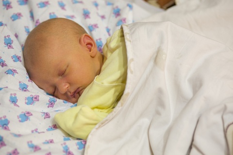
Neonatal Jaundice at Pediatric Clinic DRHC Dubai
- Jaundice is a yellow coloration of the skin and sclera and is the result of the accumulation of bilirubin.
- In most infants, hyperbilirubinemia reflects a normal transitional phenomenon, but in some infants, serum bilirubin levels may rise excessively, which can be a cause for concern because unconjugated bilirubin is neurotoxic and can cause death in newborns and lifelong neurologic sequelae in infants ( kernicterus).
- For this reason, the presence of neonatal jaundice requires immediate diagnostic evaluation.
Differential Diagnosis
- It is important to differentiate between the physiologic jaundice and other causes (pathologic) by clinical examination, laboratory, and radiologic examination.
Physiological jaundice:
- Physiological jaundice is a transient phenomenon that results from 2 phenomena. Bilirubin production is elevated because of increased breakdown of fetal erythrocytes and the higher erythrocyte mass in the neonate. Hepatic excretory capacity is low.
- Physiologic jaundice presents on the second or third day of life. Jaundice that is visible during the first 24 hours of life is likely to be non-physiologic, and further evaluation is suggested. Infants who present with jaundice after 3-4 days of life require closer monitoring.
- Physiological jaundice usually disappears within the first 2 weeks of life, so jaundice that persists for more than 2 weeks of life needs further investigation.
- In Physiologic jaundice, usually TB does not rise more than 13mg/dl in full-term baby and 15 mgldl in pre pre-term baby, so higher values need more investigations.
Etiology of Indirect Hyperbilirubinemia
Physiologic Jaundice:
- Full term newborn with peak TB 12mg/dl on the third day of life or a premature baby with peak TB 15mg/dl which occurs later (5th day).
- The peak level of physiologic jaundice may be higher in breast milk fed infant (15-17 mg/dl), which is the result of decreased fluid intake.
- Jaundice is pathologic if it is evident on the first day of life. Bilirubin level increased by more than 0.5mg/dl/hr. Peaks more than 13mg/dl in term infants; direct bilirubin more than 1mg/dl, if hepatosplenomegaly or anemia is present.
- Crigler-Najjar syndrome is a serious, rare, permanent deficiency of glucoronyl transferase and severe indirect hyperbilirubinemia. The autosomal dominant type of this disease responds to phenobarbital, while the autosomal recessive form does not respond to phenobarbital and often leads to kernicterus.
- Gilbert disease results from a mutation and manifests as a mild indirect hyperbilirubinemia.
- Breastmilk feeding is associated with unconjugated hyperbilirubinemia without evidence of hemolysis during the first 2 weeks of life, interruption of breastfeeding for 24hrs; 48hrs reduces the level of bilirubin.
- Jaundice on the first day of life is always pathologic and needs immediate attention. (Hemolysis – internal hemorrhage or infection).
- Jaundice present after 2 weeks of age is pathologic and suggests direct reacting hyperbilirubinemia.
Etiology of Direct Hyperbilirubinemia
Direct hyperbilirubninemia is not neurotoxic but signifies a serious underlying disorder:
- Cholestatsis
- CMV infection
- TORCH
- Inspissated bile from prolonged hemolysis
- Neonatal hepatitis
- Sepsis
- Inborn errors of metabolism
- Cystic fibrosis
- Biliary atresia
- Choledochal cyst
- Antitrypsin deficiency
- Alagille syndrome
- Byler syndrome
After specific diagnostic tests are performed, the supportive treatment depends on the severity of hepatocellular dysfunction and cholestasis.
Hepatocellular dysfunction predisposes the patient to complications such as bleeding, encephalopathy, and hepatorenal syndrome. Bile salt deficiency leads to fat-soluble vitamin deficiencies, Rickets, hypocalcemia, hemorrhage, and peripheral neuropathy.
Retention of bile salt may produce pruritus.
Supportive therapy involves medium-chain triglyceride and fat-soluble vitamin supplementation, cholestyramine to decrease reabsorption of bile salts from the gut.
Phenobarbital is used to increase the hepatic excretion of bile.
Definitive treatment depends on the underlying process and may involve an exclusion diet (lactose in galactosemia), surgery in extrahepatic biliary atresia or coledochal cyst, or liver transplantation.
Important Points in the Medical History
- Previous sibling with neonatal jaundice.
- Other family members with jaundice.
- Anemia – splenectomy, bile stones.
- Liver disease.
- Maternal illness.
- Maternal drug intake.
- Delay cord clamping
- Birth trauma
- Loss of stool color
- Breastfeeding
- Use of the drug
- Symptoms of hypothyroidism
- Symptoms of metabolic disease
- Exposure to total parenteral nutrition.
Physical Examination
- Neonatal jaundice first becomes visible on the face and gradually becomes visible on the trunk and extremities.
- Neurologic findings such as a change in the muscle tone, seizure in significant neonatal jaundice is danger signs and require immediate attention to prevent kernicterus.
- Hepatosplenomegaly, petechiae, and microcephaly are signs of other causes rather than physiologic jaundice, such as infections.
- Metabolic disorders require more investigations.
Lab Studies
- In infants with moderate jaundice who present on the typical second or third day of life without a history and physical examination is suggestive of a pathologic process.
- A total bilirubin level test is the only one required additional study required, which is indicated in the following studies.
- Jaundice on the first day of life.
- Infants who are anemic at birth.
- Infants who appear ill.
- Infants with TB levels are elevated enough to trigger treatment.
- Significant jaundice persists beyond the first 2 weeks of life.
- Family history of pathological jaundice.
- Hepatosplenomegaly, neurologic signs and symptoms.
The other lab tests are:
- Blood type and Rh in mother and infant.
- Direct Coombs test.
- Hg and Hct
- Blood smear
- Reticulocyte count
- Direct bilirubin
- Liver function test
- Blood culture
- Screen for IUI
- Thyroid function
- Reducing the substance in the urine
Imaging Studies
- Ultrasonography of the liver and bile ducts is warranted in an infant with laboratory or clinical signs of cholestatic disease. (hepatosplenomegaly, elevated direct bilirubin)
- Radionuclide liver scanning for uptake of (HIDA) is indicated if extrahepatic biliary atresia is suspected.
- AABR: Brainstem auditory evoked potentials should be obtained in severe neonatal jaundice to exclude sensorineural hearing loss.
Treatment:
Phototherapy, IV Ig, and exchange transfusion are the most commonly used therapeutic modalities.
- Phototherapy is the primary treatment for the neonate with unconjugated hyperbilirubinemia. Photo oxidation of the bilirubin changes some of the predominant bilirubin isomers to water-soluble isomers. The photoisomers of bilirubin are excreted in bile and urine, and they have a half-life shorter than bilirubin. Water-soluble isomers are not able to cross the blood-brain barrier, so phototherapy reduces the risk of bilirubin-induced neurotoxicity as soon as the light turns on.
- Exchange transfusions were performed when phototherapy failed to control serum bilirubin levels.
- IV Ig in an infant with Rh or AbO isoimmunization can significantly reduce the need for exchange transfusion.
- There is a special chart to show the recommendations for phototherapy and blood exchange according to TB level, age, and associated risk factors.
- The infant with phototherapy should be naked except for diapers.
- Follow up the TB level every 6-12 hrs.
- Discontinuation of phototherapy when the TB level is lower than 3mg/dl below the level that triggered the initiation of phototherapy, and follow-up tests should be obtained within 6-12 hours after discontinuation, because serum bilirubin may rebound after treatment has been discontinued.
Other Therapies:
- Infants with breastmilk jaundice: interruption of breastfeeding for 24-48 hours helps to reduce the bilirubin level.
- Prophylactic treatment of Rh mothers with Rh immunoglobulin has significantly decreased the incidence of Rh hemolytic disease.
- Surgical therapy is indicated for bile duct atresia.
Prognosis:
Prognosis is excellent if the patient receives treatment. Brain damage due to kernicterus remains a true risk. Kernicterus is a neurologic abnormality associated with hyperbilirubinemia.
Acute bilirubin encephalopathy
- First stage: Hypotonia - poor feeding.
- Second stage: Hypertonia - (backward arching of the neck), Opisthotonus
- Third stage: Hypotonia
Chronic bilirubin encephalopathy
- Extrapyramidal abnormalities: Athetosis – Chorea
- Visual abnormalities
- Auditory abnormalities
- Cognitive defect
Infants with cholestasis have a prognosis that differs according to the underlying cause. In idiopathic neonatal hepatitis, most patient recovers within 6-8 months.
In biliary atresia, the prognosis is better when the Kasai procedure is used before 3 months of age. Over 90% of patients survive 5 years after the procedure. Untreated cases die from cirrhosis before 2 years of age. In some cases of biliary hypoplasia, cirrhosis and liver failure occur before adolescence.
.png?width=281&height=59&name=bookanappointment%20(1).png)
Dubai Pediatric Clinic at Dr Rami Hamed Center provides one of the leading pediatricians in Dubai. Please call +97142798200 to book an appointment today with us!


.png?width=281&height=59&name=bookanappointment%20(1).png)
.webp?width=1080&height=1080&name=Doctor%20background%20For%20Website%20Dr.%20Manaal%2002%20(1).webp)


