Evaluation of Retina Diseases at Eye Clinic DRHC Dubai
What is Retina:
The retina, a thin layer of tissue located at the back of our eyes, plays a crucial role in capturing and transmitting visual information to our brain. Unfortunately, it is susceptible to a wide range of diseases that can impact our vision and overall eye health.
To ensure accurate diagnosis and effective treatment, it is essential to understand the significance of comprehensive evaluation. By thoroughly assessing the condition of the retina, eye care professionals can identify any abnormalities or potential issues early on. This allows for timely intervention and management strategies tailored specifically to each individual's needs.
Whether you are dealing with age-related macular degeneration, diabetic retinopathy, retinal detachment, or any other retinal condition, evaluating your retina comprehensively can make a world of difference in preserving your precious eyesight.
A comprehensive evaluation of a wide range of retina diseases:
Comprehensive eye exam:
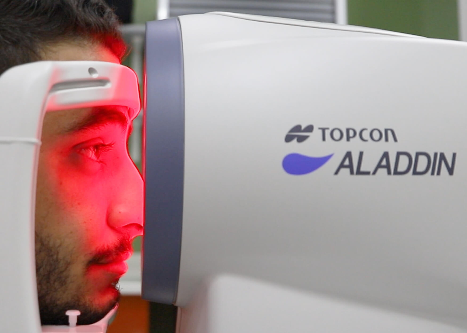
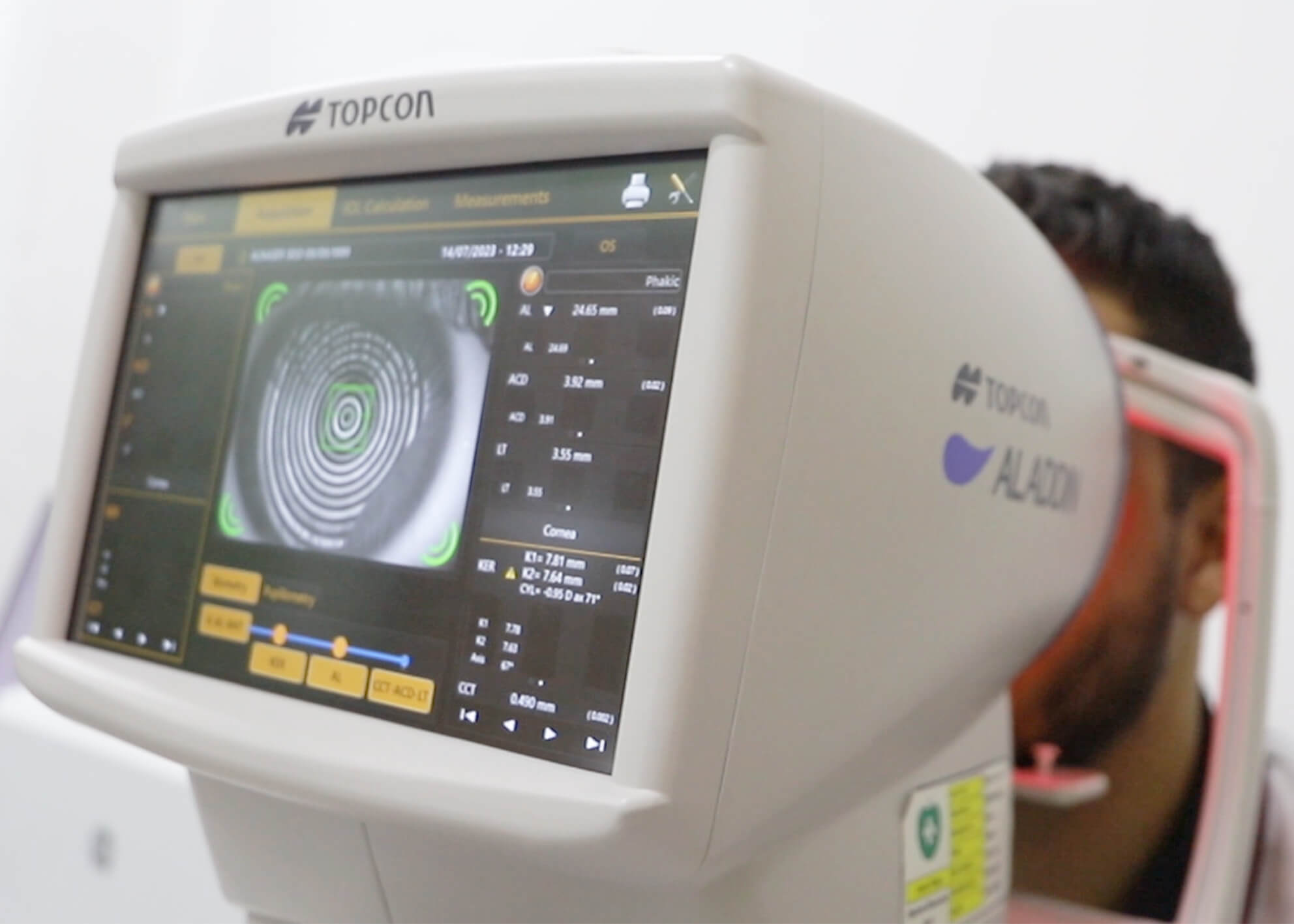
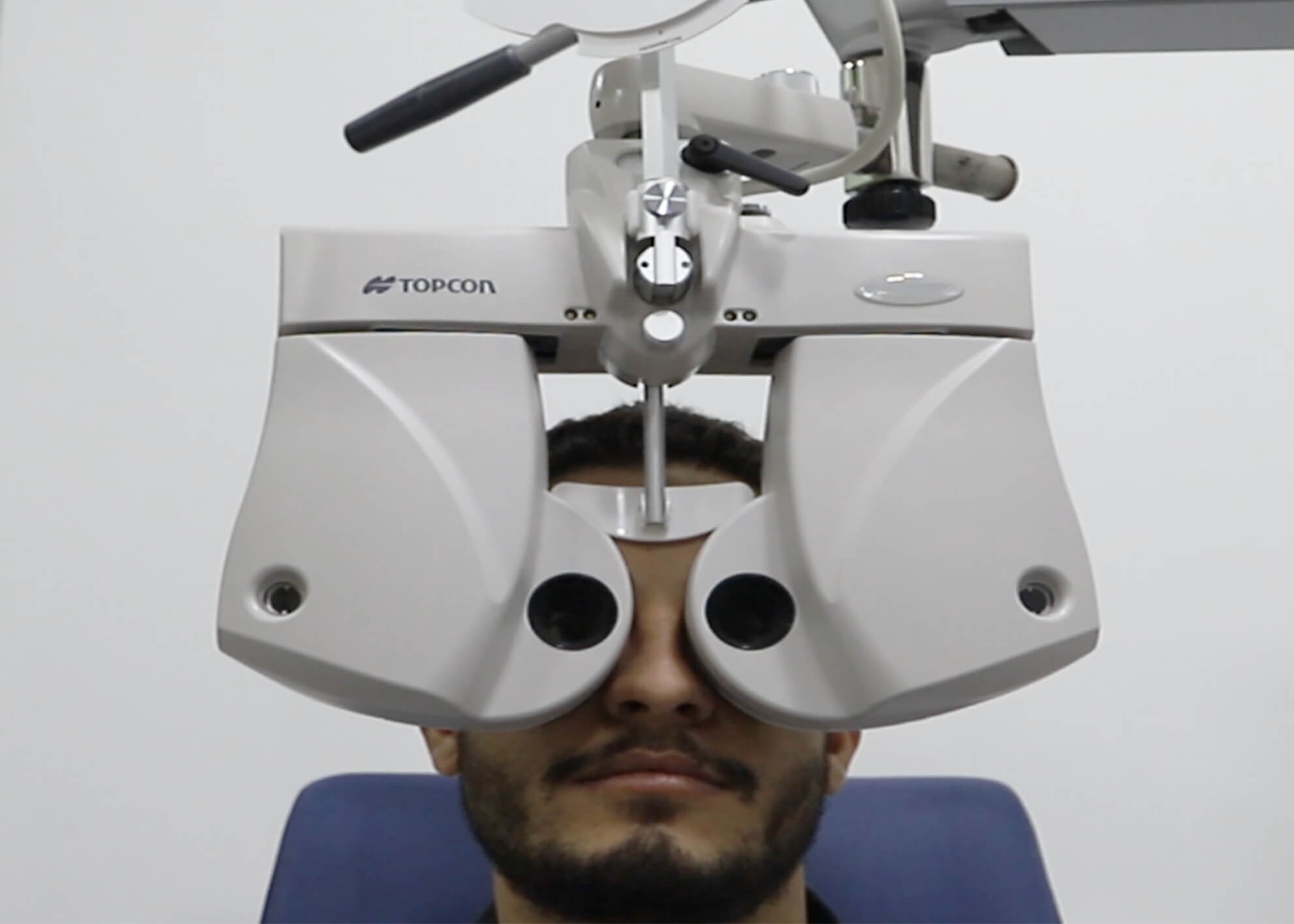
During a comprehensive eye exam, your eyes will undergo a thorough assessment, including a retinal examination. This examination allows your eye care professional to examine the back of your eyes, specifically the retina, which is essential for detecting any signs of retinal diseases or abnormalities.
A comprehensive eye exam also includes a visual acuity test, which measures how well you can see at various distances. This test helps determine if you need corrective lenses or if there are any underlying vision issues that need attention.
OCT macula:
These advanced retinal imaging techniques are revolutionizing the way we diagnose and assess eye health. By providing a detailed cross-sectional view of the macula, these scans allow for precise evaluation of retinal thickness and help detect any abnormalities or changes.
With OCT macula scans, eye care professionals can now obtain accurate and objective measurements, enabling them to make more informed decisions about patient treatment plans.
Fundus photography:
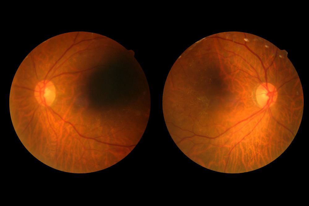
Fundus photography involves taking specialized photographs of the back of the eye, specifically the retina. These high-resolution images provide invaluable documentation of retinal conditions, aiding in accurate diagnosis and treatment planning. By capturing precise images of the retina, fundus photography enables Eye Specialists to closely monitor disease progression over time. This not only helps in assessing treatment effectiveness but also allows for early intervention when necessary.
OCT angiography:
OCTA allows us to visualize retinal blood vessels and microvasculature abnormalities with incredible detail and precision.
This technology has opened up new possibilities for early detection and monitoring of various retinal conditions, such as diabetic retinopathy, macular degeneration, and vascular occlusions.
Medical retina:
- Intravitreal injection of anti-VEGF
- Argon laser
Both are used in the management of non-surgically treated diseases like:
- Wet age-related macular degeneration (ARMD).
- Diabetic retinopathy with macular edema.
- New vessels of the retina.
- Small retinal tears.
Surgical retina:
For management of retinal diseases that need surgical intervention:
- Retinal detachment
- Giant retinal tears
- Intraocular foreign body
- Endophthalmitis
- Epiretinal membrane
- Proliferative diabetic retinopathy
Why Choose DRHC Dubai?
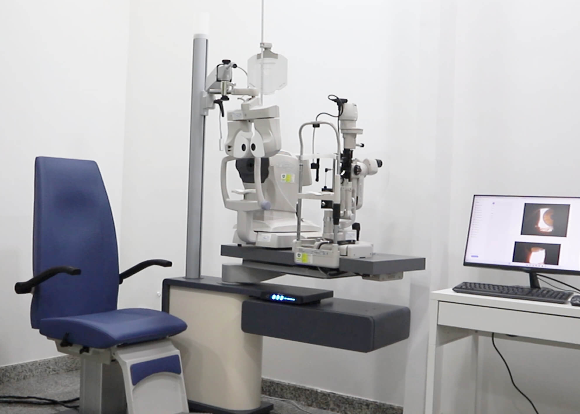
At DRHC Dubai, we prioritize your eye health with the latest diagnostic technologies and a team of expert ophthalmologists who specialize in retinal diseases. Our comprehensive evaluations ensure that we detect retinal diseases early, allowing for prompt and effective intervention. Whether you require routine monitoring or advanced surgical care, you can trust DRHC Dubai to provide the highest quality care for your retinal health.
.png?width=281&height=59&name=bookanappointment%20(1).png)
At Dr. Rami Hamed Center, our Ophthalmology department is dedicated to safeguarding your vision health through expert eye care Professionals, Renowned as one of the best eye care clinics in Dubai our Ophthalmology Specialists provide services for Cataract, Retina treatment with Laser and Refractive surgeries.


.png?width=281&height=59&name=bookanappointment%20(1).png)
