Distal Airway in Dubai at DRHC
Overview:
The distal airway includes the smaller bronchi, bronchioles, and alveoli located deep within the lungs. These structures are crucial for efficient gas exchange, allowing oxygen to enter the blood and carbon dioxide to be expelled. At DRHC Dubai, we specialize in diagnosing and treating conditions affecting the distal airway to ensure optimal respiratory health.
Anatomy of the Distal Airway:
- Bronchi: The bronchi are the large air passages that lead from the trachea to the lungs. As they branch into the lungs, they become progressively smaller.
- Bronchioles: These are smaller branches of the bronchi that lead directly to the alveoli. They are crucial for regulating airflow and distributing air to the alveoli.
- Alveoli: Tiny air sacs at the end of the bronchioles where gas exchange occurs. They are surrounded by a network of capillaries that facilitate the exchange of oxygen and carbon dioxide.
Common Distal Airway Conditions:
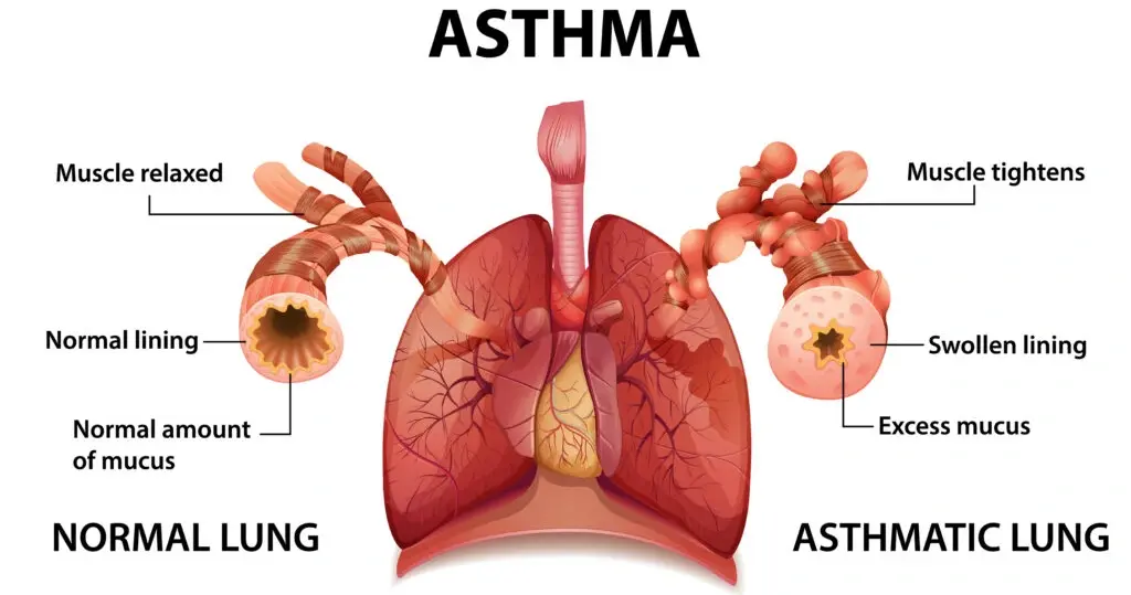
- Asthma: A chronic condition characterized by inflammation and narrowing of the bronchioles, leading to breathing difficulties.
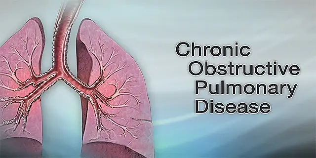
- Chronic Obstructive Pulmonary Disease (COPD): Includes chronic bronchitis and emphysema, where airflow obstruction affects the distal airways.
- Bronchiolitis: Inflammation of the bronchioles, often caused by viral infections, commonly seen in children.
- Pneumonia: Infection that inflames the alveoli, which may fill with fluid or pus, causing cough, fever, and difficulty breathing.
- Interstitial Lung Disease: A group of disorders causing scarring (fibrosis) of the lung tissue affecting the distal airways and alveoli.
Symptoms of Distal Airway Diseases:
- Shortness of breath
- Persistent cough
- Wheezing
- Chest tightness
- Reduced exercise tolerance
- Fatigue
Diagnosis:
Accurate diagnosis of distal airway conditions involves a combination of clinical evaluation, imaging studies, and functional tests, including:
- Chest X-rays and CT Scans: To visualize the structure of the lungs and detect abnormalities.
- Pulmonary Function Tests (PFTs): Measure lung capacity and airflow to assess the severity of airway obstruction.
- Bronchoscopy: A procedure using a thin, flexible tube with a camera to examine the airways.
- Sputum Tests: To identify infections or inflammation markers.
Treatment:
Treatment strategies are tailored to the specific condition and may include:
- Medications: Bronchodilators, anti-inflammatory drugs, antibiotics, and antivirals.
- Oxygen Therapy: For patients with severe respiratory impairment.
- Pulmonary Rehabilitation: A program of exercise and education to improve lung function and quality of life.
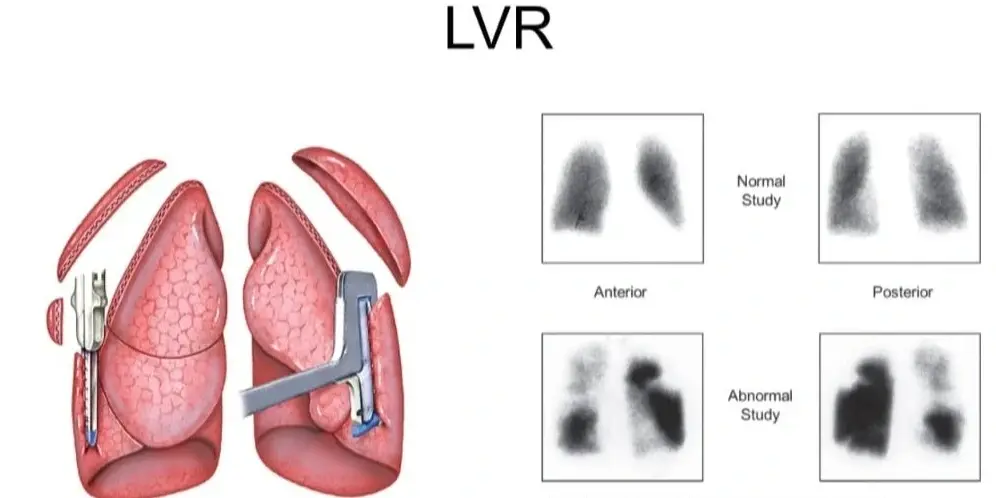
- Surgical Interventions: In severe cases, procedures like lung volume reduction surgery or lung transplantation may be considered.


.png?width=281&height=59&name=bookanappointment%20(1).png)