Pneumothorax in Dubai at DRHC
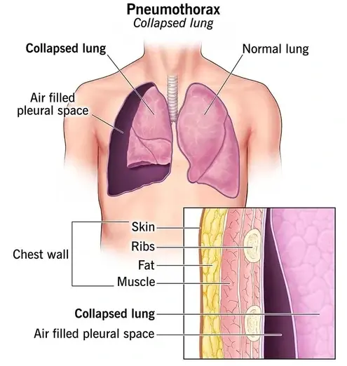
A pneumothorax occurs when air or gas accumulates in the space between the lung and the chest wall due to a hole in the lung, leading to partial or complete lung collapse. While tuberculosis was once a common cause, smoking is now the primary risk factor. Individuals with chronic obstructive pulmonary disease (COPD) are especially vulnerable to pneumothorax as their lung structure is weakened, making them more susceptible to these spontaneous air leaks.
Symptoms
Symptoms of pneumothorax can manifest during periods of rest, sleep, or wakefulness, or as a result of sudden trauma like a chest wound. A small pneumothorax may go unnoticed as it may not always present with symptoms.
For a large pneumothorax, symptoms may include:
- Intense chest pain exacerbated by coughing or deep breathing, potentially radiating to the shoulder, arm, or back
- Shortness of breath (dyspnea) or shallow breathing
- feeling of chest tightness
- Easy fatigue
- Bluish or ashen skin (cyanosis, due to lack of oxygen)
- increased heart rate (tachycardia)
Additional symptoms may involve swollen neck veins, flaring nostrils, anxiety, or low blood pressure (hypotension).
Given the varying severity of symptoms, it is not unusual for individuals to take several days to recognize the issue and seek medical attention. If you experience any symptoms of pneumothorax, it is crucial to seek immediate medical help as it can pose a life-threatening situation.
Causes
There are two primary types of pneumothorax:
Spontaneous pneumothorax can occur without a previous lung disease diagnosis or as a complication of known lung conditions such as COPD, cystic fibrosis, emphysema, asthma, tuberculosis, or whooping cough. 70% of cases happen in individuals with COPD. The cause of spontaneous pneumothorax is the presence of blebs, and small air collections between the lung and its outer surface (visceral pleura), which are typically formed during embryonic development and predominantly affect young men.
On the other hand, traumatic pneumothorax is a result of lung injury, like a gunshot, knife wound, or rib fracture. The lung can also be punctured during certain medical procedures, such as a biopsy or venous catheterization.
Occasionally, women may experience a non-traumatic pneumothorax during their menstrual period, known as catamenial pneumothorax. This unique condition occurs when endometrial tissue attaches to the thorax and forms cysts that release blood and air into the pleural space, leading to lung collapse.
Risk Factors
Men, particularly those who are tall and under the age of 40, along with individuals of Caucasian descent, face a higher risk of developing this condition.
Smoking stands out as the most significant risk factor for spontaneous pneumothorax.
Additionally, pneumothorax can have a genetic component, with up to one in 10 individuals experiencing a pneumothorax due to unknown causes having a family history of the disorder.
While the cause of pneumothorax can sometimes remain elusive, abstaining from smoking can help reduce the risk.
Diagnosis
During a physical examination, healthcare providers use a stethoscope to listen for decreased or absent breath sounds on the affected side of the lung. Additionally, they may observe a lack of expansion in the chest wall on the affected side during inhalation. Diagnostic tests that aid in confirming a pneumothorax diagnosis include Chest X-rays, Ultrasonography, Computerized tomography (CT), and Arterial blood gas testing to assess blood oxygen and carbon dioxide levels.
Treatment
In certain instances, smaller pneumothoraces may resolve on their own, but hospitalization is necessary for a large pneumothorax.
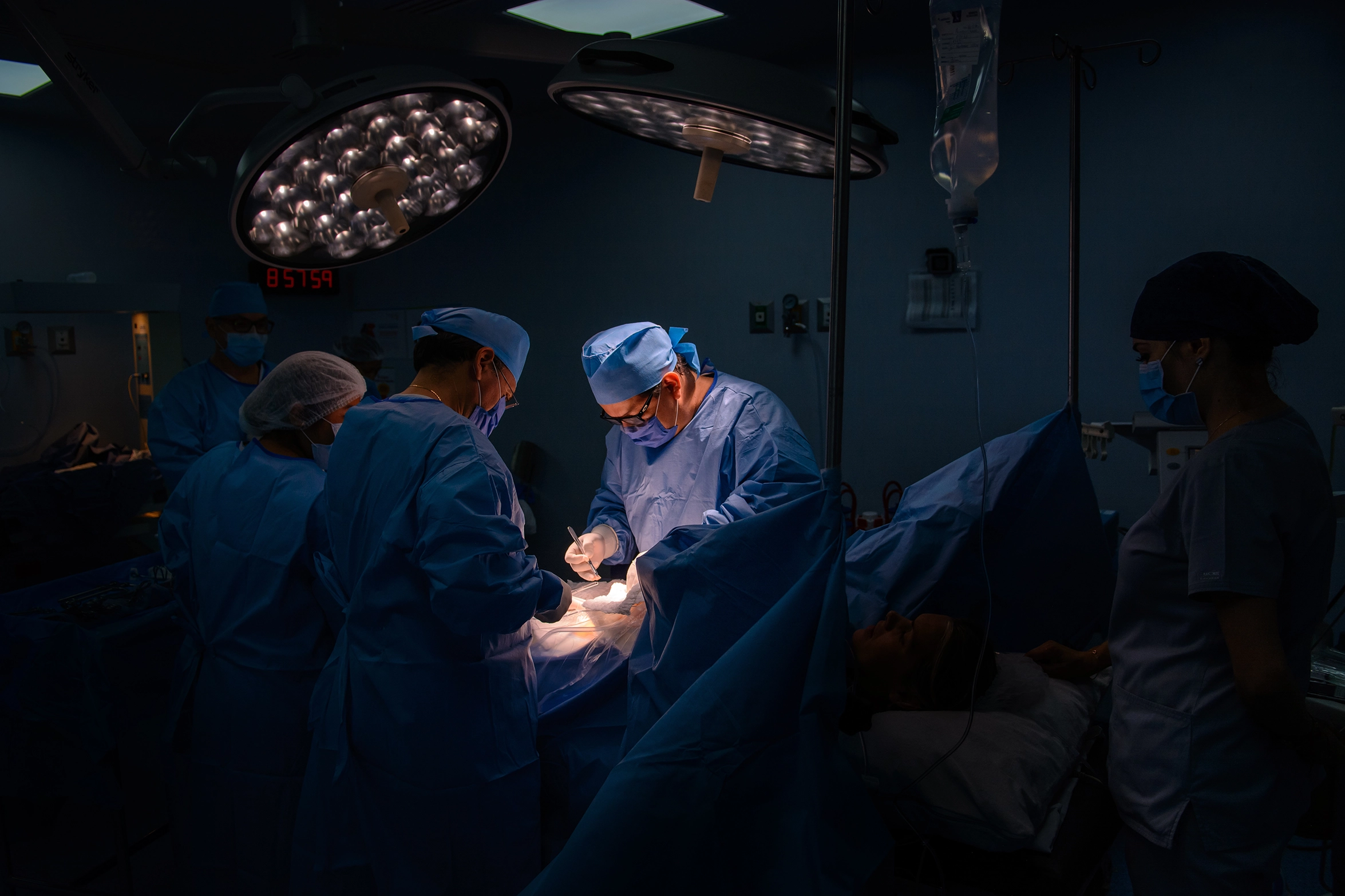
Treatment for a pneumothorax involves inserting a needle between the ribs for needle aspiration to remove air and reinflate the lung. A chest tube may also be placed and left in position for a day or longer, depending on the pneumothorax's cause, while you undergo recovery in the hospital.
If the pneumothorax reoccurs, video-assisted thoracic surgery may be recommended. The insertion of the tube or needle can cause discomfort, so pain relief may be provided through IV medication or regional anesthesia. Additionally, antibiotics might be prescribed to prevent infections. In an emergency room setting, you may be referred to a thoracic surgeon or pulmonologist for further treatment.
Recovery and Recurrence
If you have experienced a pneumothorax, it is advisable to refrain from flying until you have undergone stabilizing treatment, such as with a thoracostomy tube. Additionally, it is recommended to avoid flying or scuba diving for a period of two weeks after being discharged from the hospital following treatment. For individuals with a history of recurrent pneumothorax, it is essential to exercise caution when participating in these activities.
The risk of experiencing another pneumothorax is highest within the initial 30 days after the first incident. The risk remains elevated over the following year, with recurrence estimates ranging from 20% to as high as 60% within the first three years.
Fortunately, once a pneumothorax has healed, long-term complications are typically rare.
Click here to learn more about our surgical packages
.png?width=281&height=59&name=bookanappointment%20(1).png)
If you are in search of the best thoracic surgery in Dubai or a specialist thoracic general surgeon in Dubai call +97142798200. DRHC Dubai provides the best thoracic surgeon in Dubai and the best doctor for thoracic surgery in Dubai.


.png?width=281&height=59&name=bookanappointment%20(1).png)


